Diagram Of The Muscles In The Forearm / Forearm Muscles 13 Download Scientific Diagram - The forearm is the region of the upper limb between the elbow and the wrist.
byAdmin-
0
Diagram Of The Muscles In The Forearm / Forearm Muscles 13 Download Scientific Diagram - The forearm is the region of the upper limb between the elbow and the wrist.. The forearm is the region of the upper limb between the elbow and the wrist. The flexor pollicis longus is situated on the radial side of the forearm, lying in the same plane as the preceding. The term forearm is used in anatomy to distinguish it from the arm. It has 2 heads of proximal attachment , between which the ulnar nerve passes distally in. Learn vocabulary, terms and more with flashcards, games and other study tools.
The muscles in the posterior compartment of the forearm are commonly known as the extensor muscles. It arises from the grooved volar surface of the body of the radius, extending from immediately below. Learn vocabulary, terms and more with flashcards, games and other study tools. Diagram the movements of the humerus muscles that act on the forearm. The forearm is the region of the upper limb between the elbow and the wrist.
Muscles Of The Arm And Forearm Chart 9 from s2.thingpic.com The antibrachial or forearm muscles may be divided into a volar and a dorsal group. The forearm is a mass of some 20 different muscles. Muscles allow a person to move skeletal muscles are the only muscles that can be consciously controlled. The extrinsic hand muscles originate in the forearm and insert on structures within the hand. The term forearm is used in anatomy to distinguish it from the arm. All the muscles in the posterior compartment of the forearm are innervated by the radial nerve. Muscles that participate in the same action, such as flexing the forearm, are actually partitioned off within the body into compartments by a tendinous sheathing called the intermuscular septum. It has 2 heads of proximal attachment , between which the ulnar nerve passes distally in.
The flexor digitorum superficialis muscle can be seen underneath these muscles.
These muscles are involved of flexion and extension of the forearm at the elbow joint. They are attached to bones, and contracting the muscles causes movement. Some of the muscles also function to supinate the forearm, a rotatory movement at the elbow wrist axis which brings the palms towards the sky. Diagram the movements of the humerus muscles that act on the forearm. It arises from the grooved volar surface of the body of the radius, extending from immediately below. Start studying muscles of the forearm. The flexor pollicis longus is situated on the radial side of the forearm, lying in the same plane as the preceding. The muscle of the anterior compartment (arm in anatomical position) function as flexors while the muscles of the posterior compartment function as extensors. Pronator teres pronates the forearm, turning the hand posteriorly. This layer contains only one muscle, the flexor digitorum. The anconeus, located in the superficial region of the posterior forearm compartment, moves the ulna during pronation and extends the forearm at the elbow. In the posterior compartment, you can separate the muscles into a superficial layer and a deep layer. The forearm is the region of the upper limb between the elbow and the wrist.
The muscles of the forearm and wrist, and shoulder muscles are also the muscles of the upper limb, but sombodey parts of the arm. I've just switched over to a diagram to show you this muscle. Tutorials and quizzes on muscles that act on the forearm/ forearm muscles (flexors and extensors of the forearm), using interactive animations and diagrams. The flexor pollicis longus is situated on the radial side of the forearm, lying in the same plane as the preceding. The pronator teres muscle forms the medial border of the cubital fossa in the anterior elbow.
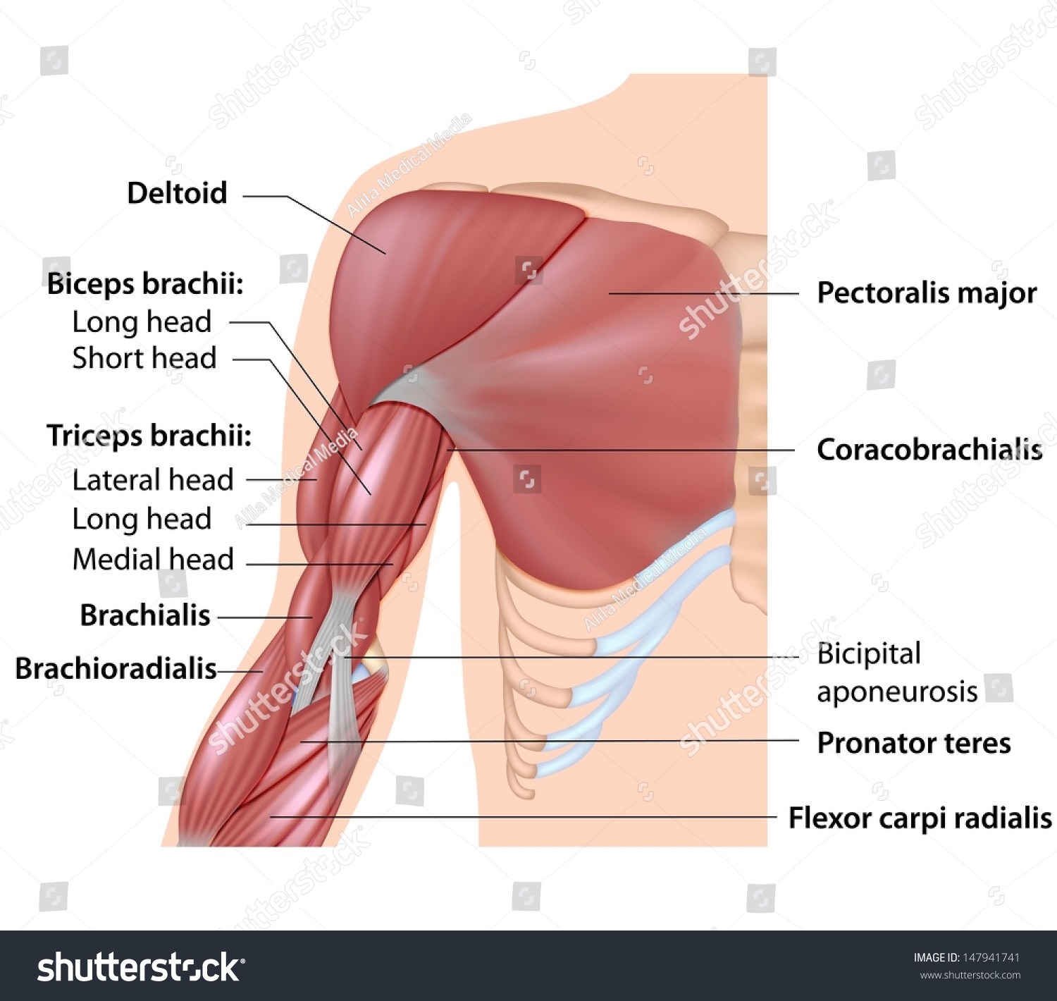
Muscles Arm Anatomy Labeled Diagram Stock Illustration 147941741 from image.shutterstock.com Tutorials and quizzes on muscles that act on the forearm/ forearm muscles (flexors and extensors of the forearm), using interactive animations and diagrams. The muscles of the upper arm are responsible for the flexion and extension of the forearm at the elbow joint. Remembering the action of each one can be quite difficult. The anterior forearm muscles are divided into 3 muscular layers ; Pronator teres pronates the forearm, turning the hand posteriorly. Muscles that move the forearm. This layer contains only one muscle, the flexor digitorum. Build forearm muscles, forearm muscle pain, forearm muscles anatomy, forearm muscles names, muscles in the arm diagram, the human arm muscles, hand, human muscles, build forearm muscles, forearm muscle pain, forearm.
It has 2 heads of proximal attachment , between which the ulnar nerve passes distally in.
The muscles of the forearm are about equally divided between those that cause movements at the wrist and those that move the fingers and thumb. The term forearm is used in anatomy to distinguish it from the arm. Diagram of the muscles of the arm in action. It is a functionally important muscle that contains two heads. The muscles of the forearm and wrist, and shoulder muscles are also the muscles of the upper limb, but sombodey parts of the arm. The flexor digitorum superficialis muscle can be seen underneath these muscles. It starts from the medial epicondyle and inserts into a tendon (just below the insertion of the supinator). Inflammation of this region caused by repetitive. The muscles of the upper arm are responsible for the flexion and extension of the forearm at the elbow joint. Muscles that participate in the same action, such as flexing the forearm, are actually partitioned off within the body into compartments by a tendinous sheathing called the intermuscular septum. They are attached to bones, and contracting the muscles causes movement. Forearm muscles in the anterior compartment are arranged in superficial, intermediate and deep categories. Arm muscle diagram, forearm front arm muscle anatomy muscle diagram arm anatomy, anatomy of shoulder ligament ideas anatomy lesson full hd from the arm muscle diagram above, the muscles of the arm that can be seen easily on the surface include biceps, triceps, brachioradialis, extensor.
The forearm is the region of the upper limb between the elbow and the wrist. Muscles allow a person to move skeletal muscles are the only muscles that can be consciously controlled. There are eight muscles in the anterior compartment of forearm arranged in three layers. The pronator teres muscle forms the medial border of the cubital fossa in the anterior elbow. Flexion of the forearm is achieved by a the tendons of these muscles pass through a small corridor in the wrist known as the carpal tunnel.
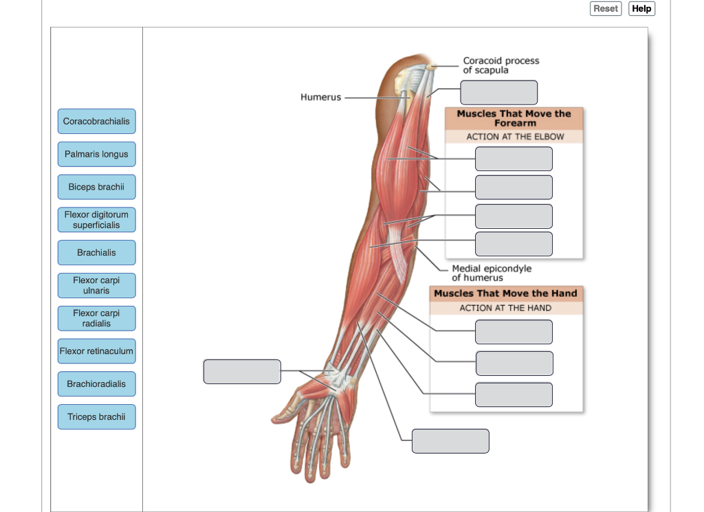
Solved Drag The Labels Onto The Diagram To Identify The M Chegg Com from media.cheggcdn.com The anconeus, located in the superficial region of the posterior forearm compartment, moves the ulna during pronation and extends the forearm at the elbow. Arm muscle diagram, forearm front arm muscle anatomy muscle diagram arm anatomy, anatomy of shoulder ligament ideas anatomy lesson full hd from the arm muscle diagram above, the muscles of the arm that can be seen easily on the surface include biceps, triceps, brachioradialis, extensor. It is a functionally important muscle that contains two heads. The muscular system consists of various types of muscle that each play a crucial role in the function of the body. This is the most medial of the superficial flexor muscles in the forearm. 4, attachment… the muscles of the back forearm. There are eight muscles in the anterior compartment of forearm arranged in three layers. The muscle of the anterior compartment (arm in anatomical position) function as flexors while the muscles of the posterior compartment function as extensors.
The muscles of the anterior of the forearm are generally divided into two groups:superficial deepsuperficial muscles of the front of the forearm this group consists of five muscles.
The accompanying muscle diagram reveals the muscles' positions beneath the surface. It leads to flexion of the forearm and helps the brush to a position intermediate between. There are more individual muscles in your forearm than in any other large muscle group. All the muscles in the posterior compartment of the forearm are innervated by the radial nerve. This is the most medial of the superficial flexor muscles in the forearm. The flexor pollicis longus is situated on the radial side of the forearm, lying in the same plane as the preceding. The muscular system consists of various types of muscle that each play a crucial role in the function of the body. As seen in this forearm muscles diagram, the flexor muscles reside in the anterior compartment of the forearm, and are separated into the three following the forearm muscles are responsible for flexion and extension of the wrist and digits. Diagram of the muscles of the arm in action. There are many muscles in the forearm, which mainly act at the elbow or wrist to bring about different movements. The antibrachial or forearm muscles may be divided into a volar and a dorsal group. Try labeling diagrams and worksheets as additional learning aids. Pronator teres pronates the forearm, turning the hand posteriorly.